A cataract is a clouding of the normally clear lens of the eye. For people who have cataracts, seeing through cloudy lenses is a bit like looking through a frosty or fogged-up window. Clouded vision caused by cataracts can make it more difficult to read, drive a car (especially at night) or see the expression on a friend’s face.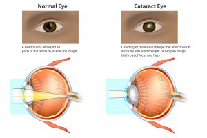
Most cataracts develop slowly and don’t disturb your eyesight early on. But with time, cataracts will eventually interfere with your vision. At first, stronger lighting and eyeglasses can help you deal with cataracts. But if impaired vision interferes with your usual activities, you might need cataract surgery. Fortunately, cataract surgery is generally a safe, effective procedure.
Symptoms
Signs and symptoms of cataracts include:
- Clouded, blurred or dim vision
- Increasing difficulty with vision at night
- Sensitivity to light and glare
- Need for brighter light for reading and other activities
- Seeing “halos” around lights
- Frequent changes in eyeglass or contact lens prescription
- Fading or yellowing of colors
- Double vision in a single eye
At first, the cloudiness in your vision caused by a cataract may affect only a small part of the eye’s lens and you may be unaware of any vision loss. As the cataract grows larger, it clouds more of your lens and distorts the light passing through the lens. This may lead to more-noticeable symptoms.
When to see a doctor
Make an appointment for an eye exam if you notice any changes in your vision. If you develop sudden vision changes, such as double vision or flashes of light, sudden eye pain, or sudden headache, see your doctor right away.
Causes
Most cataracts develop when aging or injury changes the tissue that makes up the eye’s lens. Proteins and fibers in the lens begin to break down, causing vision to become hazy or cloudy.
Some inherited genetic disorders that cause other health problems can increase your risk of cataracts. Cataracts can also be caused by other eye conditions, past eye surgery or medical conditions such as diabetes. Long-term use of steroid medications, too, can cause cataracts to develop.
How a cataract forms
A cataract is a cloudy lens. The lens is positioned behind the colored part of your eye (iris). The lens focuses light that passes into your eye, producing clear, sharp images on the retina — the light-sensitive membrane in the eye that functions like the film in a camera.
As you age, the lenses in your eyes become less flexible, less transparent and thicker. Age-related and other medical conditions cause proteins and fibers within the lenses to break down and clump together, clouding the lenses.
As the cataract continues to develop, the clouding becomes denser. A cataract scatters and blocks the light as it passes through the lens, preventing a sharply defined image from reaching your retina. As a result, your vision becomes blurred.
Cataracts generally develop in both eyes, but not always at the same rate. The cataract in one eye may be more advanced than the other, causing a difference in vision between eyes.
Types of cataracts
Cataract types include:
Cataracts affecting the center of the lens (nuclear cataracts). A nuclear cataract may at first cause more nearsightedness or even a temporary improvement in your reading vision. But with time, the lens gradually turns more densely yellow and further clouds your vision.
As the cataract slowly progresses, the lens may even turn brown. Advanced yellowing or browning of the lens can lead to difficulty distinguishing between shades of color.
- Cataracts that affect the edges of the lens (cortical cataracts). A cortical cataract begins as whitish, wedge-shaped opacities or streaks on the outer edge of the lens cortex. As it slowly progresses, the streaks extend to the center and interfere with light passing through the center of the lens.
- Cataracts that affect the back of the lens (posterior subcapsular cataracts). A posterior subcapsular cataract starts as a small, opaque area that usually forms near the back of the lens, right in the path of light. A posterior subcapsular cataract often interferes with your reading vision, reduces your vision in bright light, and causes glare or halos around lights at night. These types of cataracts tend to progress faster than other types do.
Cataracts you’re born with (congenital cataracts). Some people are born with cataracts or develop them during childhood. These cataracts may be genetic, or associated with an intrauterine infection or trauma.
These cataracts may also be due to certain conditions, such as myotonic dystrophy, galactosemia, neurofibromatosis type 2 or rubella. Congenital cataracts don’t always affect vision, but if they do, they’re usually removed soon after detection.
Risk factors
Factors that increase your risk of cataracts include:
- Increasing age
- Diabetes
- Excessive exposure to sunlight
- Smoking
- Obesity
- High blood pressure
- Previous eye injury or inflammation
- Previous eye surgery
- Prolonged use of corticosteroid medications
- Drinking excessive amounts of alcohol
Prevention
No studies have proved how to prevent cataracts or slow the progression of cataracts. But doctors think several strategies may be helpful, including:
- Have regular eye examinations. Eye examinations can help detect cataracts and other eye problems at their earliest stages. Ask your doctor how often you should have an eye examination.
- Quit smoking. Ask your doctor for suggestions about how to stop smoking. Medications, counseling and other strategies are available to help you.
- Manage other health problems. Follow your treatment plan if you have diabetes or other medical conditions that can increase your risk of cataracts.
Choose a healthy diet that includes plenty of fruits and vegetables. Adding a variety of colorful fruits and vegetables to your diet ensures that you’re getting many vitamins and nutrients. Fruits and vegetables have many antioxidants, which help maintain the health of your eyes.
Studies haven’t proved that antioxidants in pill form can prevent cataracts. But a large population study recently showed that a healthy diet rich in vitamins and minerals was associated with a reduced risk of developing cataracts. Fruits and vegetables have many proven health benefits and are a safe way to increase the amount of minerals and vitamins in your diet.
- Wear sunglasses. Ultraviolet light from the sun may contribute to the development of cataracts. Wear sunglasses that block ultraviolet B (UVB) rays when you’re outdoors.
- Reduce alcohol use. Excessive alcohol use can increase the risk of cataracts.
MANAGING AGE-RELATED MACULAR DEGENERATION (AMD)
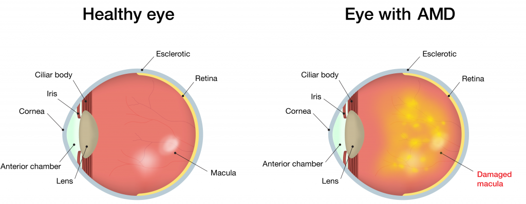
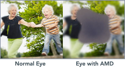
What is AMD? (AMD) Age-related macular degeneration is a condition that affects your macula – a small area in the back of your eye that is responsible for central vision. Central vision allows you to clearly see fine details, such as facial expressions or words on a page.
Older people are more at risk and the condition can worsen with age. You may not even realize you have the disease until it reaches a more advanced stage, because it can progress slowly over a long period of time.
In the most serious cases, AMD may advance with alarming speed, causing severe and irreversible central blindness within months, sometimes even weeks.
What are the symptoms of AMD?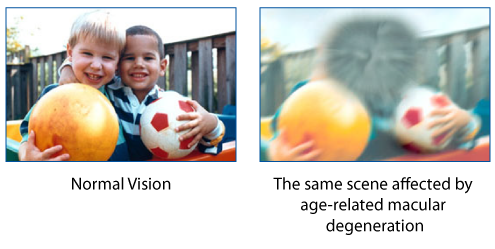
Dry AMD
Also known as early or moderate AMD, and the most common form, can result in blurred and cloudy areas of central vision. While it can alter your ability to see what’s in front of you, Dry AMD is likely to cause central vision loss.
Symptoms:
- Blurred vision
- Dark spots in front of the eyes
- Reduced sensitivity to contrast
- Difficulty adjusting from bright to dim lighting
- Difficulty reading small print
Wet AMD
Is more severe that Dry AMD and occurs when abnormal blood vessels behind the macula start to grow under the retina. They push the macula up from its normal position and leak, bleed and scar resulting in rapid central vision loss.
Symptoms:
- Large dark spots in or around the central vision
- Rapid or sudden visual loss over days or weeks
- Blindness
- Early detection and diagnosis, in either case, can help protect your sight!
What are the risk factors
Risk factors include a family history of AMD, age, light skin and blue or light-colored eyes. Other risk factors include excessive sun exposure, obesity, high blood pressure, smoking and poor diet.
Do I have it?
Even though there are several symptoms and factors, to get the best results is to book an appointment at a local Optometrist.
You can book one today at Labuschagne & Alberts Optometrists by calling: 018 468 8903 or simply come make a visit at 10 Rockwill Square Corner William & Austin Streets, Wilkoppies.
What is Glaucoma?

Glaucoma is a group of diseases that damage the eye’s optic nerve and can result in vision loss and blindness. However, with early detection and treatment, you can often protect your eyes against serious vision loss.
The optic nerve
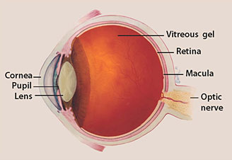
The optic nerve is a bundle of more than 1 million nerve fibers. It connects the retina to the brain. (See diagram above.) The retina is the light-sensitive tissue at the back of the eye. A healthy optic nerve is necessary for good vision.
How does the optic nerve get damaged by open-angle glaucoma?
Several large studies have shown that eye pressure is a major risk factor for optic nerve damage. In the front of the eye is a space called the anterior chamber. A clear fluid flows continuously in and out of the chamber and nourishes nearby tissues. The fluid leaves the chamber at the open angle where the cornea and iris meet. (See diagram below.) When the fluid reaches the angle, it flows through a spongy meshwork, like a drain, and leaves the eye.
In open-angle glaucoma, even though the drainage angle is “open”, the fluid passes too slowly through the meshwork drain. Since the fluid builds up, the pressure inside the eye rises to a level that may damage the optic nerve. When the optic nerve is damaged from increased pressure, open-angle glaucoma-and vision loss—may result. That’s why controlling pressure inside the eye is important.
Another risk factor for optic nerve damage relates to blood pressure. Thus, it is important to also make sure that your blood pressure is at a proper level for your body by working with your medical doctor.
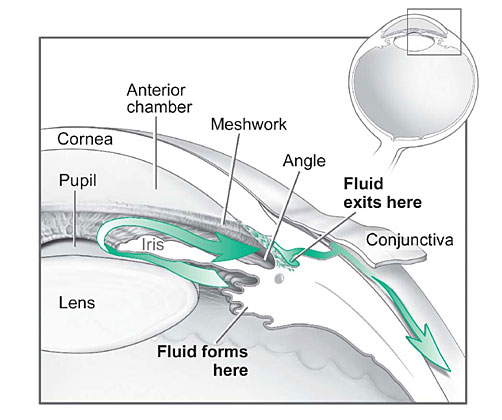
Fluid pathway is shown in teal.
Can I develop glaucoma if I have increased eye pressure?
Not necessarily. Not every person with increased eye pressure will develop glaucoma. Some people can tolerate higher levels of eye pressure better than others. Also, a certain level of eye pressure may be high for one person but normal for another.
Whether you develop glaucoma depends on the level of pressure your optic nerve can tolerate without being damaged. This level is different for each person. That’s why a comprehensive dilated eye exam is very important. It can help your eye care professional determine what level of eye pressure is normal for you.
Can I develop glaucoma without an increase in my eye pressure?
Yes. Glaucoma can develop without increased eye pressure. This form of glaucoma is called low-tension or normal-tension glaucoma. It is a type of open-angle glaucoma.
Who is at risk for open-angle glaucoma?
Anyone can develop glaucoma. Some people, listed below, are at higher risk than others:
- Everyone over age 60,
- People with a family history of glaucoma
A comprehensive dilated eye exam can reveal more risk factors, such as high eye pressure, thinness of the cornea, and abnormal optic nerve anatomy. In some people with certain combinations of these high-risk factors, medicines in the form of eyedrops reduce the risk of developing glaucoma by about half.
Glaucoma Symptoms
At first, open-angle glaucoma has no symptoms. It causes no pain. Vision stays normal. Glaucoma can develop in one or both eyes.
Without treatment, people with glaucoma will slowly lose their peripheral (side) vision. As glaucoma remains untreated, people may miss objects to the side and out of the corner of their eye. They seem to be looking through a tunnel. Over time, straight-ahead (central) vision may decrease until no vision remains.

Normal Vision.
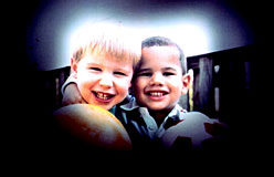
The same scene as viewed by a person with glaucoma.
How is glaucoma detected?
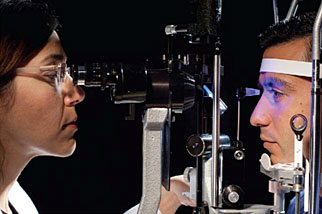
Glaucoma is detected through a comprehensive dilated eye exam that includes the following:
Visual acuity test. This eye chart test measures how well you see at various distances.
Visual field test. This test measures your peripheral (side vision). It helps your eye care professional tell if you have lost peripheral vision, a sign of glaucoma.
Dilated eye exam. In this exam, drops are placed in your eyes to widen, or dilate, the pupils. Your eye care professional uses a special magnifying lens to examine your retina and optic nerve for signs of damage and other eye problems. After the exam, your close-up vision may remain blurred for several hours.
Tonometry is the measurement of pressure inside the eye by using an instrument called a tonometer. Numbing drops may be applied to your eye for this test. A tonometer measures pressure inside the eye to detect glaucoma.
Pachymetry is the measurement of the thickness of your cornea. Your eye care professional applies a numbing drop to your eye and uses an ultrasonic wave instrument to measure the thickness of your cornea.
Can glaucoma be cured?
No. There is no cure for glaucoma. Vision lost from the disease cannot be restored.
Glaucoma Treatments
Immediate treatment for early-stage, open-angle glaucoma can delay progression of the disease. That’s why early diagnosis is very important.
Glaucoma treatments include medicines, laser trabeculoplasty, conventional surgery, or a combination of any of these. While these treatments may save remaining vision, they do not improve sight already lost from glaucoma.
Medicines. Medicines, in the form of eyedrops or pills, are the most common early treatment for glaucoma. Taken regularly, these eyedrops lower eye pressure. Some medicines cause the eye to make less fluid. Others lower pressure by helping fluid drain from the eye.
Before you begin glaucoma treatment, tell your eye care professional about other medicines and supplements that you are taking. Sometimes the drops can interfere with the way other medicines work.
Glaucoma medicines need to be taken regularly as directed by your eye care professional. Most people have no problems. However, some medicines can cause headaches or other side effects. For example, drops may cause stinging, burning, and redness in the eyes.
Many medicines are available to treat glaucoma. If you have problems with one medicine, tell your eye care professional. Treatment with a different dose or a new medicine may be possible.
Because glaucoma often has no symptoms, people may be tempted to stop taking, or may forget to take, their medicine. You need to use the drops or pills as long as they help control your eye pressure. Regular use is very important.
A tonometer measures pressure inside the eye to detect glaucoma.
Make sure your eye care professional shows you how to put the drops into your eye. For tips on using your glaucoma eyedrops, see the inside back cover of this booklet.
Laser trabeculoplasty. Laser trabeculoplasty helps fluid drain out of the eye. Your doctor may suggest this step at any time. In many cases, you will need to keep taking glaucoma medicines after this procedure.
Laser trabeculoplasty is performed in your doctor’s office or eye clinic. Before the surgery, numbing drops are applied to your eye. As you sit facing the laser machine, your doctor holds a special lens to your eye. A high-intensity beam of light is aimed through the lens and reflected onto the meshwork inside your eye. You may see flashes of bright green or red light. The laser makes several evenly spaced burns that stretch the drainage holes in the meshwork. This allows the fluid to drain better.
Like any surgery, laser surgery can cause side effects, such as inflammation. Your doctor may give you some drops to take home for any soreness or inflammation inside the eye. You will need to make several follow-up visits to have your eye pressure and eye monitored.
If you have glaucoma in both eyes, usually only one eye will be treated at a time. Laser treatments for each eye will be scheduled several days to several weeks apart.
Studies show that laser surgery can be very good at reducing the pressure in some patients. However, its effects can wear off over time. Your doctor may suggest further treatment.
Conventional surgery. Conventional surgery makes a new opening for the fluid to leave the eye. (See diagram on the next page.) Your doctor may suggest this treatment at any time. Conventional surgery often is done after medicines and laser surgery have failed to control pressure.
Conventional surgery, called trabeculectomy, is performed in an operating room. Before the surgery, you are given medicine to help you relax. Your doctor makes small injections around the eye to numb it. A small piece of tissue is removed to create a new channel for the fluid to drain from the eye. This fluid will drain between the eye tissue layers and create a blister-like “filtration bleb.”
For several weeks after the surgery, you must put drops in the eye to fight infection and inflammation. These drops will be different from those you may have been using before surgery.
Conventional surgery is performed on one eye at a time. Usually the operations are four to six weeks apart.
Conventional surgery is about 60 to 80 percent effective at lowering eye pressure. If the new drainage opening narrows, a second operation may be needed. Conventional surgery works best if you have not had previous eye surgery, such as a cataract operation.
Sometimes after conventional surgery, your vision may not be as good as it was before conventional surgery. Conventional surgery can cause side effects, including cataract, problems with the cornea, inflammation, infection inside the eye, or low eye pressure problems. If you have any of these problems, tell your doctor so a treatment plan can be developed.
What are some other forms of glaucoma and how are they treated?
Open-angle glaucoma is the most common form. Some people have other types of the disease.
In low-tension or normal-tension glaucoma, optic nerve damage and narrowed side vision occur in people with normal eye pressure. Lowering eye pressure at least 30 percent through medicines slows the disease in some people. Glaucoma may worsen in others despite low pressures.
A comprehensive medical history is important to identify other potential risk factors, such as low blood pressure, that contribute to low-tension glaucoma. If no risk factors are identified, the treatment options for low-tension glaucoma are the same as for open-angle glaucoma.
In angle-closure glaucoma, the fluid at the front of the eye cannot drain through the angle and leave the eye. The angle gets blocked by part of the iris. People with this type of glaucoma may have a sudden increase in eye pressure. Symptoms include severe pain and nausea, as well as redness of the eye and blurred vision. If you have these symptoms, you need to seek treatment immediately. This is a medical emergency. If your doctor is unavailable, go to the nearest hospital or clinic. Without treatment to restore the flow of fluid, the eye can become blind. Usually, prompt laser surgery and medicines can clear the blockage, lower eye pressure, and protect vision.
In congenital glaucoma, children are born with a defect in the angle of the eye that slows the normal drainage of fluid. These children usually have obvious symptoms, such as cloudy eyes, sensitivity to light, and excessive tearing. Conventional surgery typically is the suggested treatment, because medicines are not effective and can cause more serious side effects in infants and be difficult to administer. Surgery is safe and effective. If surgery is done promptly, these children usually have an excellent chance of having good vision.
Conventional surgery makes a new opening for the fluid to leave the eye.
Secondary glaucomas can develop as complications of other medical conditions. For example, a severe form of glaucoma is called neovascular glaucoma, and can be a result from poorly controlled diabetes or high blood pressure. Other types of glaucoma sometimes occur with cataract, certain eye tumors, or when the eye is inflamed or irritated by a condition called uveitis. Sometimes glaucoma develops after other eye surgeries or serious eye injuries. Steroid drugs used to treat eye inflammations and other diseases can trigger glaucoma in some people. There are two eye conditions known to cause secondary forms of glaucoma.
Pigmentary glaucoma occurs when pigment from the iris sheds off and blocks the meshwork, slowing fluid drainage.
Pseudoexfoliation glaucoma occurs when extra material is produced and shed off internal eye structures and blocks the meshwork, again slowing fluid drainage.
Depending on the cause of these secondary glaucomas, treatment includes medicines, laser surgery, or conventional or other glaucoma surgery.
What You Can Do
If you are being treated for glaucoma, be sure to take your glaucoma medicine every day. See your eye care professional regularly.
You also can help protect the vision of family members and friends who may be at high risk for glaucoma-African Americans over age 40; everyone over age 60, especially Mexican Americans; and people with a family history of the disease. Encourage them to have a comprehensive dilated eye exam at least once every two years. Remember that lowering eye pressure in the early stages of glaucoma slows progression of the disease and helps save vision.
Medicare covers an annual comprehensive dilated eye exam for some people at high risk for glaucoma. These people include those with diabetes, those with a family history of glaucoma, and African Americans age 50 and older.
What should I ask my eye care professional?
You can protect yourself against vision loss by working in partnership with your eye care professional. Ask questions and get the information you need to take care of yourself and your family.
What are some questions to ask?
About my eye disease or disorder…
- What is my diagnosis?
- What caused my condition?
- Can my condition be treated?
- How will this condition affect my vision now and in the future?
- Should I watch for any particular symptoms and notify you if they occur?
- Should I make any lifestyle changes?
About my treatment…
- What is the treatment for my condition?
- When will the treatment start and how long will it last?
- What are the benefits of this treatment and how successful is it?
- What are the risks and side effects associated with this treatment?
- Are there foods, medicines, or activities I should avoid while I’m on this treatment?
- If my treatment includes taking medicine, what should I do if I miss a dose?
- Are other treatments available?
About my tests…
- What kinds of tests will I have?
- What can I expect to find out from these tests?
- When will I know the results?
- Do I have to do anything special to prepare for any of the tests?
- Do these tests have any side effects or risks?
- Will I need more tests later?
Other suggestions
- If you don’t understand your eye care professional’s responses, ask questions until you do understand.
- Take notes or get a friend or family member to take notes for you. Or, bring a tape recorder to help you remember the discussion.
- Ask your eye care professional to write down his or her instructions to you.
- Ask your eye care professional for printed material about your condition.
- If you still have trouble understanding your eye care professional’s answers, ask where you can go for more information.
- Other members of your healthcare team, such as nurses and pharmacists, can be good sources of information. Talk to them, too.
Today, patients take an active role in their health care. Be an active patient about your eye care.
Loss of Vision
If you have lost some sight from glaucoma, ask your eye care professional about low vision services and devices that may help you make the most of your remaining vision. Ask for a referral to a specialist in low vision. Many community organizations and agencies offer information about low vision counseling, training, and other special services for people with visual impairments.
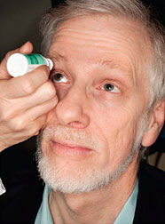
How should I use my glaucoma eyedrops?
If eyedrops have been prescribed for treating your glaucoma, you need to use them properly, as instructed by your eye care professional. Proper use of your glaucoma medication can improve the medicine’s effectiveness and reduce your risk of side effects.
To properly apply your eyedrops, follow these steps:
- Wash your hands.
- Hold the bottle upside down.
- Tilt your head back.
- Hold the bottle in one hand and place it as close as possible to the eye.
- With the other hand, pull down your lower eyelid. This forms a pocket.
- Place the prescribed number of drops into the lower eyelid pocket. If you are using more than one eyedrop, be sure to wait at least 5 minutes before applying the second eyedrop.
- Close your eye OR press the lower lid lightly with your finger for at least 1 minute. Either of these steps keeps the drops in the eye and helps prevent the drops from draining into the tear duct, which can increase your risk of side effects.
You can book one today at Labuschagne & Alberts Optometrists by calling: 018 468 8903 or simply come make a visit at 10 Rockwill Square Corner William & Austin Streets, Wilkoppies.
Keep an Eye on Ultraviolet (UV) Safety
Eye medical doctors (ophthalmologists) caution us that too much exposure to UV light raises the risks of eye diseases, including cataract, growths on the eye, and cancer. Strong exposure to snow reflection can also quickly cause painful damage called snow blindness.
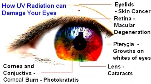
Growths on the eye, such as pterygium, can show up in our teens or twenties, especially in surfers, skiers, fishermen, farmers, or anyone who spends long hours under the mid-day sun or in the UV-intense conditions found near rivers, oceans, and mountains.
Diseases like cataract and eye cancers can take many years to develop, but each time we’re out in the sun without protection we could be adding damage that adds to our risks for these serious disorders. Babies and kids need to wear hats and sunglasses for this very reason. People of all ages should take precautions whenever they are outdoors.
Follow these tips to protect your eyes from the sun all year long:
- Sun damage to eyes can occur anytime during the year, not just in the summertime, so be sure to wear UV-blocking sunglasses and broad-brimmed hats whenever you’re outside.
- Don’t be fooled by clouds: the sun’s rays can pass through haze and thin clouds.
- Never look directly at the sun. Looking directly at the sun at any time, including during an eclipse, can lead to solar retinopathy, which is damage to the eye’s retina from solar radiation.
- Don’t forget the kids and older family members: everyone is at risk, including children and senior citizens. Protect their eyes with hats and sunglasses.
UV Light: Good in Moderation for a Good Night’s Sleep
As we sleep, our eyes enjoy continuous lubrication. During sleep the eyes also clear out irritants such as dust, allergens or smoke that may have accumulated during the day. Some research suggests that light-sensitive cells in the eye are important to our ability to regulate wake-sleep cycles. This may be more critical as we age, when more people have problems with insomnia. While it’s important that we protect our eyes from overexposure to UV light, our eyes also need minimal exposure to natural light every day to help maintain normal sleep-wake cycles.
Time Outdoors May Prevent Nearsightedness in Kids
Research shows that children who spend more time outside exposed to daylight may reduce their risk of developing nearsightedness. So not only is exercise great for eye health, but now it seem that getting that exercise while outside may be additionally beneficial. Taking your children outside to play may not only help lower their risk for nearsightedness, but will also teach them good habits for a lifetime of eye health.
We all use sunscreen to protect our skin, but don’t forget to protect your eyes as well. Summertime means more time spent outdoors, and studies show that exposure to bright sunlight may increase the risk of developing cataracts and growths on the eye, including cancer. The same risk applies when using tanning beds, so be sure to protect your eyes from indoor UV light as well. Sunlight reflected off sand and water can cause photokeratitis, the condition responsible for snow blindness, so beach- and pool-goers take note.
“UV radiation, whether from natural sunlight or indoor artificial rays, can damage the eye’s surface tissues as well as the cornea and lens,” said Michael Kutryb, MD, an ophthalmologist in Edgewater, Fla., and clinical correspondent for the American Academy of Ophthalmology. “Unfortunately, many people are unaware of the dangers UV light can pose. By wearing UV-blocking sunglasses, you can enjoy the summer safely while lowering your risk for potentially blinding eye diseases and tumors.” It is important to start wearing proper eye protection at an early age to protect your eyes from years of ultraviolet exposure.
According to a national Sun Safety Survey conducted by the American Academy of Ophthalmology, only about half of people who wear sunglasses say they check the UV rating before buying.The good news is that you can easily protect yourself. In order to be eye smart in the sun, the American Academy of Ophthalmology recommends the following:
Wear sunglasses labeled “100% UV protection”: Use only glasses that block both UV-A and UV-B rays and that are labeled either UV400 or 100% UV protection.
- Choose wraparound styles so that the sun’s rays can’t enter from the side.
- If you wear UV-blocking contact lenses, you’ll still need sunglasses.
Wear a hat along with your sunglasses; broad-brimmed hats are best.
Remember the kids: It’s best to keep children out of direct sunlight during the middle of the day. Make sure they wear sunglasses and hats whenever they are in the sun.
Know that clouds don’t block UV light: The sun’s rays can pass through haze and clouds. Sun damage to the eyes can occur any time of year, not just in summer.
Be extra careful in UV-intense conditions: Sunlight is strongest mid-day to early afternoon, at higher altitudes, and when reflected off of water, ice or snow.
By embracing these simple tips you and your family can enjoy the summer sun safely while protecting your vision.
If you feel like you need an eye test you can book one today at Labuschagne & Alberts Optometrists by calling: 018 468 8903 or simply come make a visit at 10 Rockwill Square Corner William & Austin Streets, Wilkoppies.
Dry eye syndrome is a chronic and typically progressive condition. Depending on its cause and severity, it may not be completely curable. But in most cases, dry eyes can be managed successfully, usually resulting in noticeably greater eye comfort, fewer dry eye symptoms, and sometimes sharper vision as well.
Because dry eye disease can have a number of causes, a variety of treatment approaches are used. Successful treatment of dry eyes requires that you are willing to follow your doctor’s recommendations and that you use the products he or she recommends consistently and as frequently as directed.
Artificial Tears
For mild cases of dry eyes caused by computer use, reading, schoolwork and other situational causes, the best dry eye treatment may simply be frequent use of artificial tears or other lubricating eye drops.
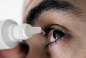
Artificial tears usually are the first step in dry eye treatment.
There are many brands of artificial tears that are available without a prescription. The challenge with using artificial tears is not lack of product availability — it’s the confusing number of brands and formulations available to choose from.
Artificial tears and other over-the-counter (OTC) lubricating eye drops are available in a wide variety of ingredients and viscosity (“thickness”).
Artificial tears with low viscosity are “light” and watery. They often provide quick relief with little or no blurring of your vision when you apply them. But often their soothing effect is very short-lived, and sometimes you must use these drops very frequently to get adequate dry eye relief.
On the other hand, artificial tears that have a high viscosity are more gel-like and can provide longer-lasting lubrication. But typically these drops cause significant blurring of your vision for several minutes immediately after you apply them. For this reason, these drops often are not a good choice for use during your work day or when you need immediate clear vision for tasks such as driving. Instead, high-viscosity artificial tears are recommended only for bedtime use.
Also, the ingredients in certain brands of artificial tears may determine which type of dry eye condition they are better suited for. For example, one brand might work better for aqueous-deficiency dry eyes, while another brand may be more effective for an evaporative dry eye condition.
If your eye doctor recommends that you use one or more brands or formulations of artificial tears, be sure to follow the directions he or she gives you concerning when and how often you use the drops. Also, do not substitute different brands from those your eye doctor recommends. Using a different brand or multiple brands of artificial tears will make it difficult to assess the success of the dry eye treatment your doctor recommended.
Punctal Plugs
Punctal plugs are sometimes used in dry eye treatment to help tears remain on the surface of the eye longer.
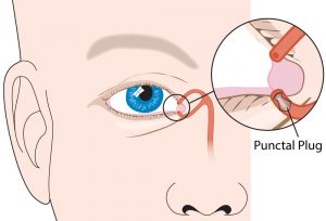
This drawing shows the lacrimal glands and tear ducts. A lacrimal plug (or punctal plug) has been inserted into the lower tear duct to keep the eye’s moisture from draining away too quickly. Image: Oasis Medical, Inc.
A punctal plug is a small, sterile device that is inserted into one of the small openings (puncta) of tear drainage ducts that are located in the inner corner of the upper and lower eyelids.
After these openings have been plugged, tears can no longer drain away from the eye through these ducts. In this way the tear film stays intact longer on the surface of the eye, relieving dry eye symptoms.
So where do the tears go? Usually they will simply evaporate from the eye surface without symptoms. But if insertion of punctal plugs causes the eyes to “water,” one or more of the plugs can be removed.
If you would like to learn more or book an appointment at Labuschagne & Alberts Optometrists simply call: 018 468 8903 or come make a visit at 10 Rockwill Square Corner William & Austin Streets, Wilkoppies.
Eye Allergy Diagnosis
Eye allergies develop when the body’s immune system becomes sensitized and overreacts to something in the environment that typically causes no problem in most people. An allergic reaction can occur when that “something” (called an allergen) comes in contact with antibodies attached to the mast cells in your eyes; the cells respond by releasing histamine and other substances or chemicals that cause tiny blood vessels to leak and the eyes to become itchy, red and watery.
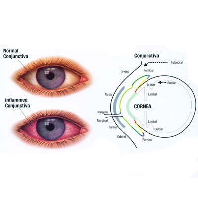
Eye allergies share symptoms with some diseases of the eye, making accurate diagnosis imperative. The symptoms of eye allergy can range from mildly annoying redness to inflammation severe enough to impair vision. If symptoms persist or over-the-counter remedies do not bring relief, see an GP or Optometrist who will review your medical history and symptoms and conduct tests that can reveal an eye allergy.
Those tests may include an examination with a microscope, which will show swollen blood vessels on the surface of the eye. In addition, your doctor may test for a certain type of white blood cell that shows up on areas of the eye affected by allergies. This involves gently scraping the conjunctiva (the inner lining of the eyelid) and seeing if those cells are found.
The primary types of eye allergy are seasonal or perennial allergic conjunctivitis, vernal keratoconjunctivitis, atopic keratoconjunctivitis, contact allergic conjunctivitis and giant papillary conjunctivitis.
Seasonal and perennial allergic conjunctivitis
Seasonal allergic conjunctivitis (SAC) is by far the most common type of eye allergy. Patients experience symptoms in spring, summer or fall, depending on the type of plant pollens in the air. Typical symptoms include:
- Itching
- Redness
- Burning
- Clear, watery discharge
People with SAC may have chronic dark circles (known as allergic shiners) under their eyes. The eyelids may be puffy, and bright lights may be bothersome. SAC symptoms often accompany the runny nose, sneezing and nasal congestion associated with hay fever and other seasonal allergies. The itching may be so bothersome that patients rub their eyes frequently, making symptoms worse and potentially causing infection.
Perennial allergic conjunctivitis (PAC), as its name implies, occurs year-round. Symptoms are the same as with SAC, but tend to be milder. They are caused by reactions to dust mites, mold, pet dander or other household allergens, rather than pollen.
Vernal keratoconjunctivitis
Vernal keratoconjunctivitis is a more serious eye allergy than SAC or PAC. While it can occur year-round, symptoms may worsen seasonally. It primarily occurs in boys and young men; about 75 percent of patients also have eczema or asthma. Symptoms include:
- Itching
- Significant tearing and production of thick mucus
- The feeling of having something in the eye (foreign body sensation)
- Aversion to light (photophobia)
If left untreated, vernal keratoconjunctivitis can impair vision.
Atopic keratoconjunctivitis
This type of allergy primarily affects older patients – mostly men with a history of allergic dermatitis. Symptoms of atopic keratoconjunctivitis can occur year-round and are similar to those of vernal keratoconjunctivitis:
- Severe itching
- Burning
- Redness
- Significant production of thick mucus that, after sleep, may cause the eyelids to stick together
If left untreated, atopic keratoconjunctivitis can result in scarring of the cornea and its delicate membrane.
Contact allergic conjunctivitis
This can result from irritation by contact lenses or by the proteins from tears that bind to the surface of the lens. Symptoms include:
- Redness
- Itching
- Mucous discharge
- Lens discomfort
Giant papillary conjunctivitis
Associated with wearing contact lenses, giant papillary conjunctivitis is a severe form of contact allergic conjunctivitis in which individual fluid sacs, or papules, form in the upper lining of the inner eyelid. Symptoms include:
- Itching
- Puffiness
- Tearing
- Mucous discharge
- Blurred vision
- Poor tolerance for wearing contact lenses
- Foreign body sensation
The first approach in managing seasonal or perennial forms of eye allergy should be to avoid the allergens that trigger your symptoms.
Outdoor exposure:
- Stay indoors as much as possible when pollen counts are at their peak, usually during the midmorning and early evening, and when wind is blowing pollens around.
- Avoid using window fans that can draw pollens and molds into the house.
- Wear glasses or sunglasses when outdoors to minimize the amount of pollen getting into your eyes.
- Try not to rub your eyes, which will irritate them and could make your condition worse.
Indoor exposure:
- Keep windows closed, and use air conditioning in your car and home. Air conditioning units should be kept clean.
- Reduce exposure to dust mites, especially in the bedroom. Use “mite-proof” covers for pillows, comforters and duvets, and mattresses and box springs. Wash your bedding frequently, using hot water (at least 130 degrees Fahrenheit).
- To limit exposure to mold, keep the humidity in your home low (between 30 and 50 percent) and clean your bathrooms, kitchen and basement regularly. Use a dehumidifier, especially in the basement and in other damp, humid places, and empty and clean it often. If mold is visible, clean it with detergent and a 5 percent bleach solution.
- Clean floors with a damp rag or mop, rather than dry-dusting or sweeping.
Exposure to pets:
- Wash your hands immediately after petting any animals. Wash your clothes after visiting friends with pets.
- If you are allergic to a household pet, keep it out of your home as much as possible. If the pet must be inside, keep it out of the bedroom so you are not exposed to animal allergens while you sleep.
- Close the air ducts to your bedroom if you have forced-air or central heating or cooling. Replace carpeting with hardwood, tile or linoleum, all of which are easier to keep dander-free.
Many allergens that trigger eye allergies are airborne, so you can’t always avoid them. Discuss your symptoms with your GP or Optometrist to determine which treatment options are right for you.
Nonprescription (over-the-counter, or OTC) eyedrops and oral medications are commonly used for short-term relief of some symptoms. They may not relieve all symptoms, and prolonged use of some OTC eyedrops may actually cause your condition to worsen.
Prescription eyedrops and oral medications also are used to treat eye allergies. The prescription drops provide both short- and long-term targeted relief of eye allergy symptoms. Your GP or Optometrist can help determine which treatments are best for you.
Children can be treated with both OTC and prescription eyedrops and medications. Artificial tears are safe and can be used at any age. Some eyedrops, such as antihistamines and mast cell stabilizers, can be used in children 3 and older. Any treatment should be discussed with your child’s physician.
OTC eyedrops and medications
- Tear substitutes: Artificial tears can temporarily wash allergens from the eye and also moisten the eyes, which often become dry when red and irritated. These drops, which can be refrigerated to provide additional soothing and comfort, are safe and can be used as often as needed.
- Decongestants: OTC decongestant eyedrops reduce the redness associated with eye allergies by narrowing the blood vessels in the eye. (Note: These should not be used by anyone with glaucoma.) They are available with a decongestant only or with a decongestant and an OTC antihistamine, which provides additional relief from itching. Because the drops are weak, they must be used frequently (four to six times a day).Do not use these OTC decongestant eyedrops for more than two to three days. Prolonged use can create a “rebound effect” – increased swelling and redness that may last even after discontinuing the drops. You may be familiar with this if you have used decongestant nasal sprays for more than three days and your nose has become even more congested than it was before.
- Oral antihistamines: While oral antihistamines can be mildly effective in relieving the itching associated with eye allergies, they may cause dry eyes and potentially worsen eye allergy symptoms. Also, some OTC versions of these medications can cause side effects such as sedation, excitability, dizziness or disturbed coordination.
Prescription eyedrops and medications
- Antihistamine eyedrops: These can reduce the itching, redness and swelling associated with eye allergies. Although these drops provide quick relief, the effect may last only a few hours, and some must be used four times a day.
- Mast cell stabilizer eyedrops: These prevent the release of histamine and other substances that cause allergy symptoms. To prevent itching, the drops must be used before you’re exposed to an allergen.
- Antihistamine and mast cell stabilizer eyedrops: Some of the newest eyedrops have both an antihistamine and a mast cell stabilizer to treat and prevent eye allergies. They are used twice a day and provide quick, long-lasting relief of itching, redness, tearing and burning.
- NSAID eyedrops: Nonsteroidal anti-inflammatory drugs (NSAIDs) are available in eyedrops to relieve itching. These drops may cause stinging or burning when applied and may need to be used four times a day.
- Corticosteroid eyedrops: These can help treat chronic, severe eye allergy symptoms such as itching, redness and swelling. Long-term treatment with steroids (more than two weeks) should be done only under the supervision of an ophthalmologist; side effects of continued use include a risk of infection, glaucoma and cataracts.
- Nonsedating oral antihistamines: Prescription antihistamines can be mildly effective in relieving the itching associated with eye allergies. While they do not have the same sedating side effects as OTC antihistamines, these medications can cause dry eyes and worsen symptoms.
- Allergy shots (immunotherapy): Allergy shots work by improving an individual’s tolerance to the substance that causes an allergic reaction. Tiny amounts of the allergen are injected with gradually increasing doses over time. The treatment takes several months to achieve maximum results, and you may still be required to use medications to alleviate symptoms.
If you would like to learn more or book an appointment at Labuschagne & Alberts Optometrists simply call: 018 468 8903 or come make a visit at 10 Rockwill Square Corner William & Austin Streets, Wilkoppies.